Position statement

Retinopathy of prematurity: Strategies to optimize screening and management
Posted: Oct 23, 2026
Principal author(s)
Gabriel Altit MD, Marc Beltempo MD, Philippe Lamer NNP, Nasrin Najm-Tehrani MB BCh MSc; Canadian Paediatric Society, Fetus and Newborn Committee
Abstract
This updated Canadian Paediatric Society (CPS) statement on retinopathy of prematurity (ROP) addresses key areas where understanding and treatment of this proliferative disorder affecting preterm infants have advanced since 2016, with emphasis on timely and appropriate screening based on postmenstrual age. Recent practice changes in neonatology, the emergence of a revised classification system (ICROP3), and advances in treatment have improved screening recommendations for ROP. Evidence-based criteria and algorithms that balance early detection with minimizing unnecessary exams anchor screening guidance. Advances in treatment, particularly anti-vascular endothelial growth factor therapies, are discussed alongside traditional laser treatments. The importance of comprehensive follow-up to optimize visual outcomes is also highlighted. A structured approach to screening and treatment that uses both pharmacological and non-pharmacological methods to enhance infant comfort during ROP examinations is recommended.
Keywords: Anti-VEGF therapy; Retinopathy of prematurity; ROP screening
Retinopathy of prematurity (ROP) is a proliferative retinal vascular disorder in preterm infants that can lead to visual impairment or blindness. In Canada, 2022 data show that 50% of infants born before 31 weeks develop ROP, with 15% of those developing ROP requiring treatment with laser and/or anti-vascular endothelial growth factor (VEGF) injections (AVIs) [1]. Since the 2016 Canadian Paediatric Society (CPS) statement [2], several changes have occurred, including updated saturation targets, improved survival of high-risk infants, new tele-ophthalmology tools, and growing support for a more selective screening approach[3]. Advances in classification, treatment, and emerging evidence warrant this statement update, which outlines revised screening criteria, new therapies, and key topics not previously addressed.
Definition and classification
ROP results from incomplete retinal vascular development in preterm infants, especially those born <31 weeks. ROP is driven by factors such as hypoxia/hyperoxia, VEGF dysregulation, retinal immaturity, inflammation, and poor postnatal growth. The International Classification of ROP (ICROP) provides standardized terminology and was most recently updated in 2021 (ICROP3) [4]. ROP is classified by retinal zone, stage (severity at the vascular-avascular junction), extent (clock hours), and the presence of plus or pre-plus disease (Figure 1 and Table 1).
Recommendation:
- ICROP3 is recommended for classifying, illustrating, and documenting retinal findings.
Figure 1. Right and left eye retina
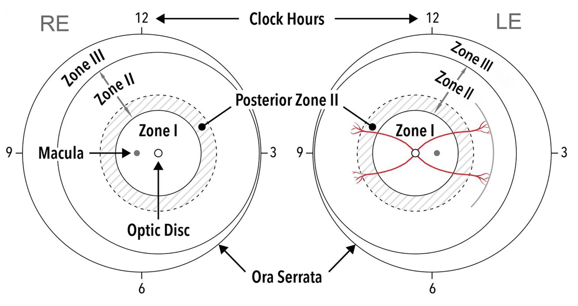
- Schematic of the RE (right eye) and LE (left eye) illustrating zone boundaries and clock-hour sectors used to describe ROP location and extent. Solid circles indicate zones I to III; dotted circles show posterior zone II (2-disc diameters beyond zone I).
- Within the macula: The fovea is a small depression located at the centre of the macula and represents the area of highest visual acuity and cone density. The fovea is responsible for sharp vision needed for reading and fine visual tasks.
- The ora serrata is the peripheral junction between the neurosensory retina and the non-photosensitive ciliary epithelium. The ora serrata represents the anterior limit of the retina.
Category | Description | |
Zones of ROP |
| |
Plus and pre-plus disease |
| |
Stages of acute disease |
| |
Retinal detachment |
| |
Extent of disease | Measured in 12 clock-hour sectors | |
Regression | Patterns of ROP regression (spontaneous or post-treatment): Complete or incomplete, with documentation of peripheral avascular retina. | |
Reactivation | ROP reactivation (new ROP lesion or vascular changes) occurring after treatment. | |
Type 1 ROP5 |
| |
Type 2 ROP5 |
| |
Aggressive ROP | Severe and rapidly progressing ROP in zone I or posterior zone II that can also extend beyond posterior retina. May occur in larger preterm infants, especially in areas of the world where health care resources are limited. | |
Source: Chiang MF, Quinn GE, Fielder AR, et al. International classification of retinopathy of prematurity, 3rd edn. Ophthalmology 2021;128(10):e51-e68. Adapted with permission.
Whom to screen
In addition to birth weight (BW) and gestational age (GA), postnatal weight has been explored to refine ROP screening criteria [6]-[9]. Weight gain-based models offer good sensitivity and negative likelihood ratios for identifying infants at low risk for ROP requiring treatment [10]. Tools like DIGIROP-Birth [11] and DIGIROP-Screen [12] use birth characteristics to guide screening decisions.
The Canadian SCREENROP study [3] analyzed 4888 infants (from 2014 to 2016) and found that screening infants with BW <1200 g and/or <30 weeks GA effectively identified those at risk of clinically significant ROP, while reducing unnecessary exams. Data from the Canadian Neonatal Network (CNN) from 2016 to 2022 showed that fewer than three infants (out of 1341 with known eye examination results) born between 30+0 and 30+6 weeks GA and weighing over 1200 grams required ROP treatment. Due to confidentiality policies, exact numbers are not disclosed. Notably, these rare cases involved infants with uncertain GA, later estimated to be 31 weeks. Canadian level-three neonatal intensive care unit (NICU) data also confirmed that no infant born at 31 weeks and >1200 grams required ROP treatment. These findings support a potential revision of the 2016 CPS screening criteria (<30 weeks and <1200 grams). However, the analysis does not include infants assessed or treated exclusively in level two units or outpatient clinics. Individuals with a history of prematurity or ROP—particularly those with severe or treated disease (especially when exposed to AVI)—should be made aware of potential long-term ocular sequelae and the importance of continued eye care into adulthood.
Recommendations regarding screening eligibility:
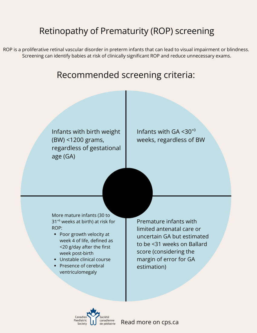
- The CPS now recommends adjusting ROP screening criteria as follows:
- Infants with BW <1200 grams, regardless of GA
- Infants with GA <30+0 weeks, regardless of BW
- Premature infants with limited antenatal care or uncertain GA but estimated to be <31 weeks on Ballard score (considering the margin of error for GA estimation)
- More mature infants (30 to 31+6 weeks at birth) at risk for ROP:
- Poor growth velocity at week 4 of life, defined as <20 g/day after the first week of postnatal age ([weight at day 28 – weight at day 7] / 21 days <20 g/day), independent of BW or size relative to GA,
- Unstable clinical course (those exposed to inotropes, vasopressors, or both),
OR - Presence of cerebral ventriculomegaly, defined as anterior horn width or ventricular index >2 standard deviations above the mean for GA [13].
Schedule of screening examination
Scheduling should aim to identify infants needing treatment in a timely manner while minimizing exams in low-risk infants. Since ROP develops later in most immature infants, the timing of first exam should be based on postmenstrual age. Suggested schedules (Table 2) are informed by clinical trials [14]-[16], with limited data in infants <26 weeks suggesting that significant ROP typically appears after 31 weeks post-menstrual age (PMA; refers to GA at birth + chronological age after birth) [17][18]. A baseline exam before treatment helps assess disease progression and allows families and care teams to prepare. Based on supporting CNN data from 2016/2022 and the absence of new evidence, the timing of initial screening remains unchanged from the 2016 CPS guidance [2]. Follow-up exam intervals should be determined by the ophthalmologist, with guidance in Table 3, based on previous recommendations [2].
Birth GA, weeks | Age at initial examination, weeks | |
Postmenstrual age in weeks | Chronological age in weeks | |
22 | 31 | 9 |
23 | 31 | 8 |
24 | 31 | 7 |
25 | 31 | 6 |
26 | 31 | 5 |
27 | 31 | 4 |
28 | 32 | 4 |
29 | 33 | 4 |
≥30 | ≥34 | 4 |
VEGF Vascular endothelial growth factor
Source: Retinopathy of prematurity: Strategies to optimize screening and management.
© Canadian Paediatric Society, 2026. Also available at www.cps.ca
Tele-ophthalmology
Tele-ophthalmology ROP screening generally refers to the acquisition of retinal imaging via a retinal camera and remote review of the images for diagnosis by an ophthalmologist in a referral centre. This allows infants to be cared for in a level two centre that may be closer to home, an approach which has been successfully implemented in different settings [19]-[21]. One limitation is that the camera may not capture the far peripheral retina near the ora serrata precisely (Figure 1). While this strategy appears to be cost-effective in North American contexts, further study is needed to assess its cost-effectiveness across diverse health care settings in Canada [22][23].
Recommendations regarding ROP screening:
- Schedule initial screening at time of admission and/or at 4 weeks of age to ensure no delays or missed eligible infants. Follow-up screening(s) should be determined by the ophthalmologist, in consultation with neonatal staff, with timing based on the risk of progression to a treatable stage. Scheduled follow-ups must be communicated to families in advance.
- ROP screening must be conducted by trained professionals capable of providing comfort measures, administering dilating drops, maintaining sterility, and performing assessments, all with the support of a paediatric ophthalmologist, either in-person or via telemedicine.
- Results of all screening exams must be documented and communicated to parents and the infant’s care team in a timely manner.
- A regional tele-ophthalmology ROP screening program may be considered, provided the following resources are ensured [20][21]:
- Availability of frontline health care professionals (e.g., neonatologists, paediatricians, non-paediatric ophthalmologists, or other disciplines) trained to capture the retinal images at the appropriate screening timepoints with suitable equipment [24],
- Dedicated resources allocated for training professionals, acquiring and maintaining equipment, and setting up a system of safe data transfer, and
- A secure framework for identification, timing of evaluation, and sustained access, with a built-in backup system featuring two trained individuals in each centre.
Performance of screening and promoting comfort
Retinal screenings should be conducted with pupillary dilation and sterile instruments. Dilating drops should be used judiciously due to cardiac (bradycardia, tachycardia, hypertension) and gastrointestinal (feeding intolerance, regurgitations, delayed gastric emptying) side effects. Lower dosage of phenylephrine 0.5% and cyclopentolate 0.1% are effective for dilating the neonatal pupil [25] adequately while possibly reducing adverse effects. A single examination may suffice, but only if it conclusively shows that the retina is fully vascularized in both eyes.
The CPS Managing pain in newborns statement recommends sweet-tasting solutions, non-nutritive sucking, containment, and parental presence or voice as comfort measures during ROP screening. Studies have shown that acetaminophen [26] or intranasal fentanyl [27] may also help reduce exam-related pain, although there is insufficient data to recommend systematic administration of these agents. Music therapy, such as recorded maternal singing and heartbeat, may also ease infant distress and support recovery [28]. Clinicians should be aware that ROP exams can cause transient cardiorespiratory instability (e.g., bradycardia, apnea, desaturations) and delayed gastric emptying [29]. Involving a parent or additional staff member to provide comfort alongside the examiner—creating a “four-handed care” approach—may further reduce infant stress. Four-handed care refers to the additional caregiver (parent, nurse, or staff member) providing comfort, containment, and non-pharmacological soothing.
Recommendations for comfort during ROP screening and care:
- Topical anesthetic, mydriatic, and cycloplegic drops should be administered before ROP screening or treatment to dilate pupils and enhance comfort. Where available, lower-concentration formulations may be preferred to minimize systemic side effects [30][31].
- Non-pharmacological measures, including but not limited to parental presence, bundling or swaddling, a pacifier “sweetened” with breast milk or sucrose, “four-handed care”, and music therapy should also be used. Skin-to-skin contact, gentle touching or holding in the moments surrounding exam can be considered, depending on the comfort of the family and examiner. The CPS statement Skin-to-skin care (SSC) for term and preterm infants states that SSC alleviates procedural pain in preterm infants, especially when combined with breastfeeding.
- Feeding infants in the NICU an hour before ROP examination, as opposed to withholding feeds for 2 or more hours, may help reduce stress and improve physiological stability during the procedure [32].
- Acetaminophen (10–15 mg/kg, administered 30 to 60 minutes before ROP examination) may provide mild pain relief, though evidence for recommending this is limited. Acetaminophen use should be reserved for infants who have previously shown significant distress during exams, rather than administered routinely.
- Low-dose intranasal fentanyl (1 mcg/kg) may be considered in select cases—such as in infants prone to instability or expected to experience significant discomfort during the exam.
Preventing ROP
ROP prevention begins in the NICU with strategies such as optimizing nutrition and postnatal growth, maintaining recommended oxygen saturation targets between 90% and 95% [33][34], and minimizing intermittent hypoxemic episodes [35]. Evidence suggests that both early hyperoxia and later fluctuations in oxygenation are associated with increased ROP risk. Emerging studies support individualized oxygen strategies in established ROP to reduce progression [36][37]. ROP prevention must also consider modifiable systemic factors including delivery room stabilization, optimal oxygen targeting, and consistent practices across units, in addition to nutritional and respiratory strategies.
Treating ROP
ROP treatment’s goal is to halt abnormal vascular proliferation that leads to retinal detachment and blindness. Postnatal extrauterine hyperoxic state suppresses retinal VEGF, resulting in halted vessel growth and avascular retina. Subsequent ischemia then triggers excess VEGF, leading to pathological angiogenesis in ROP. Laser therapy reduces VEGF production by ablating the avascular retina, while AVIs directly block VEGF activity. Treatment decisions depend on disease progression, ROP type, infant stability, available resources, and may require transfer to a level 3 centre to access expertise. Practitioners should balance potential benefits and risks—including systemic absorption and reactivation—and collaborate closely with families.
Laser and AVI
Timely, near-confluent laser therapy remains highly effective for severe acute ROP, promoting regression and preventing retinal detachment [38]. AVIs have been used for nearly two decades as primary therapy, rescue treatment after failed laser, or as adjuncts in severe cases. Multiple trials since 2011 have supported their effectiveness in acute ROP treatment [39]-[41].
Potential AVI benefits
Laser irreversibly ablates the peripheral avascular retina, whereas AVI allows regression of ROP without tissue destruction and supports continued peripheral vascularization, even in severe posterior disease. As AVI drug levels decline, there remains a risk of ROP reactivation. Ten-year follow-up data from the CRYO-ROP study showed a small but measurable reduction in peripheral visual fields among patients treated with laser compared with those whose ROP resolved spontaneously [42]. Although long-term comparative data on visual fields between laser and AVI are lacking, the ability of the latter to allow retinal vascularization is promising. Studies suggest lower rates of myopia [43] and fewer binocular function disruptions [44] in AVI-treated infants compared with those treated with laser. Laser typically requires general anesthesia with intubation, while AVI can often be administered at the bedside using topical and mild systemic analgesia (intranasal fentanyl, subcutaneous morphine, acetaminophen, or intravenous low-dose analgesia). These factors, including anesthesia risks, should be discussed with families when determining the best treatment approach, particularly in cases where GA, transport, or intubation may pose additional risks.
Potential complications of AVI
Risks of intravitreal injections include potential injury to intraocular structures and endophthalmitis, which can result in permanent vision loss. AVI should only be performed by expert ophthalmologists, using strict aseptic technique and with a thorough understanding of the unique ocular anatomy of premature infants.
Surveillance post-AVI
Infants treated with AVI require a clear, coordinated follow-up plan, often with more frequent and prolonged surveillance than those treated with laser alone. Responsibility for monitoring ROP reactivation or persistent avascular retina (PAR)—and for initiating further treatment—rests with the ophthalmologist(s) with ROP expertise. A recent Canadian study of 238 eyes treated with bevacizumab (an AVI) reported a 27% retreatment rate, mainly for PAR [45]. The finding highlights the need for individualized follow-up and, in some cases, need for laser to reduce long-term risks, emphasizing tailored surveillance and shared decision-making. If discharge from the NICU leads to transfer of care, this responsibility must be formally handed over to an ophthalmologist with the necessary expertise and access to appropriate infrastructure. Long-term follow-up should include comprehensive assessments of visual function, including refraction, ocular motility, binocular vision, and retinal structure—potentially requiring general anesthesia or retinal angiography in select cases.
Potential risks to infants
Given the lack of definitive data on the systemic effects of AVI in premature infants, it is essential that caregivers are informed that the long-term safety data are limited. Several studies have shown that AVI can reduce circulating VEGF levels, raising concerns about potential impacts on developing organs [46]. These risks should be clearly communicated, and ongoing research is actively investigating this important area.
Recommendations regarding treatment:
- Treatment within 48 to 72 hours is recommended for “type 1 ROP” (occasionally termed “high-risk pre-threshold ROP”) and aggressive ROP (Table 1). Type 2 ROP requires close individualized monitoring.
- Surgical intervention may be considered to promote retinal reattachment and preserve vision when ROP progresses to stage 4 or 5 (partial or total detachment).
- Engage families in treatment decisions that include discussions of benefits, risks, and uncertainties around use of laser and AVI, while considering local resources and potential long-term effects.
Stopping criteria and post-discharge follow-up
Premature infants with regressed ROP—whether spontaneous or after treatment—remain at risk for ophthalmic sequelae such as refractive errors, amblyopia, and strabismus. These conditions are preventable and treatable causes of long-term visual impairment and can be identified through regular follow-up with eye care professionals experienced in assessing infants and young children.
Screening for acute ROP can be discontinued in infants who never developed ROP or whose ROP regressed spontaneously without treatment. However, a comprehensive eye exam at 6 to 9 months corrected age remains important for all preterm infants who met criteria for ROP screening [47][48]. If no abnormalities are detected, further follow-up should follow routine paediatric care guidelines, including vision and eye health assessment around 3 years corrected age. Long-term sequelae of ROP may include late retinal detachment, persistent peripheral avascular retina, macular abnormalities, ongoing retinal vascular changes, and glaucoma. Therefore, long-term follow-up beyond infancy should be planned in collaboration with ophthalmologists, with periodic assessments throughout early childhood and school age.
Recommendations regarding parental education, follow-up, and general care considerations:
- Explain ROP, with its risks and outcomes, including the potential for visual impairment or blindness, to families and caregivers. Emphasize the importance of regular screening, timely intervention, and adherence to follow-up for families, caregivers, and the primary health care team.
- Preventing missed ROP follow-up appointments requires early and ongoing education of caregivers and health care providers regarding the importance of adhering to post-discharge ophthalmological care.
- When acute ROP has regressed, routine retinal examinations with scleral depression are no longer necessary unless otherwise clinically indicated.
- After NICU discharge, the clinician performing the final ROP examination should establish and clearly communicate a follow-up eye care plan to both caregivers and NICU staff.
- A comprehensive ophthalmological assessment is recommended at 6 to 9 months corrected age for all preterm infants who met criteria for ROP screening, including age-appropriate visual acuity assessment, ocular motility evaluation, cycloplegic refraction, and dilated fundus examination.
- Units performing ROP screening should participate in quality assurance initiatives related to ROP prevention, screening, treatment practices, and personnel competency.
- Support national registries that continuously monitor ROP practices and outcomes because approximately 50% of neonates born at <31 weeks develop some stage of ROP, and 7% require treatment.
- Implementation of these recommendations requires local adaptation based on available resources, including access to trained personnel and paediatric ophthalmologists, and should incorporate communication strategies that support and inform families while minimizing anxiety.
Acknowledgements
We would like to thank Dr. Ann Jefferies for reviewing the content of this statement and providing suggestions and adjustments. We would also like to thank the Canadian Neonatal Network for providing data from 2016 to 2022. This statement was reviewed by the Neonatal-Perinatal Medicine Section Executive and the Community Paediatrics and Drug Therapy Committees of the Canadian Paediatric Society. It was also reviewed by representatives of the Canadian Association of Neonatal Nurses (CANN), the Canadian Association for Pediatric Ophthalmology and Strabismus (CAPOS), and the Canadian Retina Society (CRS).
CANADIAN PAEDIATRIC SOCIETY FETUS AND NEWBORN COMMITTEE (2024-2025)
Members: Souvik Mitra MD MSc (Chair), Michael Narvey MD (Past Chair), Sidd Thakore MD (Board Representative), Gabriel Altit MD, Nicole Radziminski MD, Emer Finan MB, Mireille Guillot MD, Meghan Gibbery (Resident Member), Sarah Lawrence MD
Liaisons: William Ehman MD (College of Family Physicians of Canada), Chantal Nelson (Public Health Agency of Canada), Eric Eichenwald MD (American Academy of Pediatrics, Committee on Fetus & Newborn), R. Douglas Wilson MD (The Society of Obstetricians and Gynaecologists of Canada), Isabelle Milette
RN, MScN, NNP (Canadian Association of Neonatal Nurses), Deepak Manhas MD (CPS Neonatal-Perinatal Medicine Section)
Principal authors: Gabriel Altit MD, Marc Beltempo MD, Philippe Lamer NNP, Nasrin Najm-Tehrani MB BCh MSc
Potential Conflict of Interest
Dr. Altit is supported by FRQS Junior 1 Clinician Scientist and has also received peer-reviewed funding support from the Montreal Children’s Hospital Foundation and Foundation for Pediatric Research. The NeoCardioLab website, the platform of Dr. Altit, has received an unrestricted grant from Mallinckrodt pharmaceuticals. No other disclosures were reported. All authors have submitted the ICMJE Form for Disclosure of Potential Conflicts of Interest.
Funding
There is no funding to declare.
References
- Canadian Neonatal Network/Le Réseau Néonatal Canadien. Annual Report/Rapport Annuel; 2022. Presentation 23: Retinopathy of prematurity treatments: GA <33 weeks (Accessed June 23, 2026).
- Jefferies AL; Canadian Paediatric Society, Fetus and Newborn Committee. Retinopathy of prematurity: An update on screening and management. Paediatr Child Health 2016;21(2):101-8. doi: 10.1093/pch/21.2.101
- Sabri K, Shivananda S, Farrokhyar F, et al. Refining evidence-based retinopathy of prematurity screening guidelines: The SCREENROP study. Paediatr Child Health 2019;25(7):455-66. doi: 10.1093/pch/pxz085. Corrigendum in Paediatr Child Health 2021;26(7):444. doi: 10.1093/pch/pxab065
- Chiang MF, Quinn GE, Fielder AR, et al. International classification of retinopathy of prematurity, third edition. Ophthalmology 2021;128(10):e51-e68. doi: 10.1016/j.ophtha.2021.05.031
- Early Treatment for Retinopathy of Prematurity Cooperative Group. Revised indications for the treatment of retinopathy of prematurity: Results of the early treatment for retinopathy of prematurity randomized trial. Arch Ophthalmol 2003;121(12):1684-94. doi: 10.1001/archopht.121.12.1684
- Binenbaum G, Bell EF, Donohue P, et al. Development of modified screening criteria for retinopathy of prematurity: Primary results from the Postnatal Growth and Retinopathy of Prematurity Study. JAMA Ophthalmol 2018;136(9):1034-40. doi: 10.1001/jamaophthalmol.2018.2753
- Quinn GE, Ying GS, Bell EF, et al. Incidence and early course of retinopathy of prematurity: Secondary analysis of the Postnatal Growth and Retinopathy of Prematurity (G-ROP) Study. JAMA Ophthalmol 2018;136(12):1383-9. doi: 10.1001/jamaophthalmol.2018.4290
- Binenbaum G. Retinopathy of prematurity screening—Asking the proper question. JAMA Ophthalmol 2023:141(8):725-6. doi: 10.1001/jamaophthalmol.2023.2782
- Zupancic JAF, Ying GS, de Alba Campomanes A, Tomlinson LA, Binenbaum G. Evaluation of the economic impact of modified screening criteria for retinopathy of prematurity from the Postnatal Growth and ROP (G-ROP) study. J Perinatol 2020;40(7):1100-8. doi: 10.1038/s41372-020-0605-5
- Athikarisamy S, Desai S, Patole S, Rao S, Simmer K, Lam GC. The use of postnatal weight gain algorithms to predict severe or type 1 retinopathy of prematurity: A systematic review and meta-analysis. JAMA Netw Open 2021;4(11):e2135879. doi: 10.1001/jamanetworkopen.2021.35879
- Pivodic A, Hård A-L, Löfqvist C, et al. Individual risk prediction for sight-threatening retinopathy of prematurity using birth characteristics. JAMA Ophthalmol 2020;138(1):21-9. doi: 10.1001/jamaophthalmol.2019.4502
- Pivodic A, Johansson H, Smith LEH, et al. Development and validation of a new clinical decision support tool to optimize screening for retinopathy of prematurity. Br J Ophthalmol 2022;106(11):1573-80. doi: 10.1136/bjophthalmol-2020-318719
- El-Dib M, Limbrick DD, Inder T, et al. Management of post-hemorrhagic ventricular dilatation in the infant born preterm. J Pediatr 2020;226:16-27.e13. doi: 10.1016/j.jpeds.2020.07.079
- Reynolds JD, Dobson V, Quinn GE, et al. Evidence-based screening criteria for retinopathy of prematurity: Natural history data from the CRYO-ROP and LIGHT-ROP studies. Arch Ophthalmol 2002;120(11):1470-6. doi: 10.1001/archopht.120.11.1470
- Cryotherapy for Retinopathy of Prematurity Cooperative Group. Multicenter trial of cryotherapy for retinopathy of prematurity. Three-month outcome. Arch Ophthalmol 1990;108(2):195-204. doi: 10.1001/archopht.1990.01070040047029
- Kennedy KA, Wrage LA, Higgins RD, et al. Evaluating retinopathy of prematurity screening guidelines for 24- to 27-week gestational age infants. J Perinatol 2014;34(4):311-8. doi: 10.1038/jp.2014.12
- Austeng D, Källen KBM, Hellström A, Tornqvist K, Holmström GE. Natural history of retinopathy of prematurity in infants born before 27 weeks’ gestation in Sweden. Arch Ophthalmol 2010;128(10):1289-94. doi: 10.1001/archophthalmol.2010.234
- Austeng D, Källen KB, Hellström A, et al. Screening for retinopathy of prematurity in infants born before 27 weeks’ gestation in Sweden. Arch Ophthalmol 2011;129(2):167-72. doi: 10.1001/archophthalmol.2010.346
- Solebo AL, Teoh L, Sargent J, Rahi JS; British Childhood Visual Impairment and Blindness Study Interest Group. Avoidable childhood blindness in a high-income country: Findings from the British Childhood Visual Impairment and Blindness Study 2. Br J Ophthalmol 2023;107(12):1787-92. doi: 10.1136/bjo-2022-321718
- Abdul Aziz AA, Isaac M, Tehrani NN. Using telemedicine to screen for retinopathy of prematurity. CMAJ 2014186(13):1012-4. doi: 10.1503/cmaj.131357
- Tehrani NN. Telemedicine screening for retinopathy of prematurity (ROP): Three years experience in Ontario, Canada. Investig Ophthalmol Visual Sci 2012;53(14):5881
- Jackson KM, Scott KE, Graff Zivin J, et al. Cost-utility analysis of telemedicine and ophthalmoscopy for retinopathy of prematurity management. Arch Ophthalmol 2008;126(4):493-9. doi: 10.1001/archopht.126.4.493
- Isaac M, Isaranuwatchai W, Tehrani NN. Cost analysis of remote telemedicine screening for retinopathy of prematurity. Can J Ophthalmol 2018;53(2):162-7. doi: 10.1016/j.jcjo.2017.08.018
- Quinn GE, Ying GS, Daniel E, et al. Validity of a telemedicine system for the evaluation of acute-phase retinopathy of prematurity. JAMA Ophthalmol 2014;132(10):1178-84. doi: 10.1001/jamaophthalmol.2014.1604
- Kremer LJ, Medlicott N, Sime MJ, et al. Low dose or very low dose phenylephrine and cyclopentolate microdrops for retinopathy of prematurity eye examinations (The Little Eye Drop Study): A randomised controlled non-inferiority trial. Arch Dis Child Fetal Neonatal Ed 2023;108(4):380-6. doi: 10.1136/archdischild-2022-324929
- Kabataş EU, Dursun A, Beken S, Dilli D, Zenciroğlu A, Okumuş N. Efficacy of single dose oral paracetamol in reducing pain during examination for retinopathy of prematurity: A blinded randomized controlled trial. Indian J Pediatr 2016;83(1):22-6. doi: 10.1007/s12098-015-1765-8
- Naik A, D’Lima A, Sreekumar K, Silveira MP. Efficacy of expressed breast milk alone or in combination with paracetamol in reducing pain during ROP screening: A randomized controlled trial. J Clin Neonatol 2021;10(2):73-8. doi: 10.4103/jcn.jcn_198_20
- Corrigan MJ, Keeler JR, Miller HD, Ben Khallouq BA, Fowler SB. Music therapy and retinopathy of prematurity screening: Using recorded maternal singing and heartbeat for post exam recovery. J Perinatol 2020;40(12):1780-8. doi: 10.1038/s41372-020-0719-9
- Tan JBC, Dunbar J, Hopper A, Wilson CG, Angeles DM. Differential effects of the retinopathy of prematurity exam on the physiology of premature infants. J Perinatol 2019;39(5):708-16. doi: 10.1038/s41372-019-0331-z
- Alselaimy R, Al Tawil L, Abouammoh MA. Anesthesia in retinopathy of prematurity. Saudi J Ophthalmol 2022;36(3):251-9. doi: 10.4103/sjopt.sjopt_229_21
- Marsh VA, Young WO, Dunaway KK, et al. Efficacy of topical anesthetics to reduce pain in premature infants during eye examinations for retinopathy of prematurity. Ann Pharmacother 2005;39(5):829-33. doi: 10.1345/aph.1E476
- Strube YNJ, Bakal JA, Arthur BW. Relationship between feeding schedules and gastric distress during retinopathy of prematurity screening eye examinations. J AAPOS 2010;14(4):334-9. doi: 10.1016/j.jaapos.2010.05.008
- Askie LM, Darlow BA, Davis PG, et al. Effects of targeting lower versus higher arterial oxygen saturations on death or disability in preterm infants. Cochrane Database Syst Rev 2017;4(4):CD011190. doi: 10.1002/14651858.CD011190.pub2
- Moreton RBR, Fleck BW, Fielder AR, et al. The effect of oxygen saturation targeting on retinal blood vessel growth using retinal image data from the BOOST-II UK Trial. Eye (Lond) 2016;30(4):577-81. doi: 10.1038/eye.2015.280
- Di Fiore JM, Bloom JN, Orge F, et al. A higher incidence of intermittent hypoxemic episodes is associated with severe retinopathy of prematurity. J Pediatr 2010;157(1):69-73. doi: 10.1016/j.jpeds.2010.01.046
- Cayabyab R, Arora V, Wertheimer F, Durand M, Ramanathan R. Graded oxygen saturation targets and retinopathy of prematurity in extremely preterm infants. Pediatr Res 2016;80(3):401-6. doi: 10.1038/pr.2016.98
- Minturn R, Hartigan K, Vegunta S, et al. Efficacy of supplemental oxygen in reducing the need for laser or intravitreal bevacizumab in preterm infants with stage 2 retinopathy of prematurity. BMC Ophthalmol 2024;24(1):220. doi: 10.1186/s12886-024-03483-w
- Emami S, Isaac M, Mireskandari K, Tehrani NN. Laser treatment for retinopathy of prematurity: A decade since ETROP. Ophthalmology 2019;126(4):639-41. doi: 10.1016/j.ophtha.2018.12.012
- Mintz-Hittner HA, Kennedy KA, Chuang AZ; BEAT-ROP Cooperative Group. Efficacy of intravitreal bevacizumab for stage 3+ retinopathy of prematurity. N Engl J Med 2011;364(7):603-15. doi: 10.1056/NEJMoa1007374
- Stahl A, Lepore D, Fielder A, et al. Ranibizumab versus laser therapy for the treatment of very low birthweight infants with retinopathy of prematurity (RAINBOW): An open-label randomised controlled trial. Lancet 2019;394(10208):1551-9. doi: 10.1016/S0140-6736(19)31344-3
- Stahl A, Sukgen EA, Wu WC, et al. Effect of intravitreal aflibercept vs laser photocoagulation on treatment success of retinopathy of prematurity: The FIREFLEYE randomized clinical trial. JAMA 2022;328(4):348-59.
- Quinn GE, Dobson V, Hardy RJ, Tung B, Phelps DL, Palmer EA. Visual fields measured with double-arc perimetry in eyes with threshold retinopathy of prematurity from the cryotherapy for retinopathy of prematurity trial. The CRYO-Retinopathy of Prematurity Cooperative Group. Ophthalmology 1996;103(9):1432-7. doi: 10.1016/s0161-6420(96)30487-9
- Geloneck MM, Chuang AZ, Clark WL, et al. Refractive outcomes following bevacizumab monotherapy compared with conventional laser treatment: A randomized clinical trial. JAMA Ophthalmol 2014132(11):1327-33. doi: 10.1001/jamaophthalmol.2014.2772
- Manuchian S, Mireskandari K, Tehrani NN, Isaac M, Robitaille JM. Binocularity outcomes following treatment for retinopathy of prematurity. Can J Ophthalmol 2021;56(3):179-83. doi: 10.1016/j.jcjo.2020.10.007
- Isaac M, Mireskandari K, Kertes PJ, Tehrani NN. Follow-up algorithm after bevacizumab treatment for retinopathy of prematurity. Ophthalmol Retina 2025;9(7):709-11. doi: 10.1016/j.oret.2025.02.010
- Chawla D, Darlow BA. Anti-vascular endothelial growth factor preparations in the treatment of retinopathy of prematurity: Balancing risks and benefits. Indian Pediatr 2016;53(Suppl 2):s129-s136.
- Fierson WM; American Academy of Pediatrics, Section on Ophthalmology, American Academy of Ophthalmology, American Association for Pediatric Ophthalmology and Strabismus, American Association of Certified Othoptists. Screening examination of premature infants for retinopathy of prematurity. Pediatrics 2018;142(6):e20283061. doi: 10.1542/peds.2018-3061
- Hutchinson AK, Morse CL, Hercinovic A, et al. Pediatric eye evaluations preferred practice pattern. Ophthalmology 2023:130(3):P222-P270. doi: 10.1016/j.ophtha.2022.10.030
Disclaimer: The recommendations in this position statement do not indicate an exclusive course of treatment or procedure to be followed. Variations, taking into account individual circumstances, may be appropriate. Internet addresses are current at time of publication.